Written by:

Senior Principal Scientist, BioScience Renal, Cardiovascular, Renal and Metabolism, BioPharmaceuticals R&D

Executive Director, Head of Biosciences Renal, BioPharmaceuticals R&D
Chronic kidney disease (CKD) is a serious, progressive condition and under-recognised public health problem, affecting nearly 840 million people worldwide.1 More than 9 million people living with CKD have progressed to kidney failure, requiring dialysis or a kidney transplant, and by 2040, CKD is expected to become the world’s 5th leading cause of mortality.2,3,4
By observing the intricacies of a complete physiological system, we can gain a better understanding of dysfunction in disease, and this may be the key to transforming care in CKD. Kidneys are complex, composed of approximately 20-40 different cell types, arranged within an elaborate architectural matrix. CKD is characterised by the engagement of multiple molecular pathways, connections, and complex pathophysiology, all of which contribute to the challenge of developing new therapies for this condition.
Previously, researchers would use static, one or two cell models, add investigational compounds and then examine any potential interaction. However, these standard, non-fluidic, 2D cell-based models have not been able to adequately reflect the 3D complexity of the kidney environment (including microfluid flow, shear stress and cell signalling). As such, results are not always a faithful representation of what happens in the body.
A 3D culture system (e.g., kidney microphysiological systems or kidney-on-chips) offers a more realistic ‘real time’ window into what goes on in CKD. Kidney microphysiological systems (MPS) allow different cell types to be co-cultured, enabling cellular interactions and signalling to be investigated in both healthy and diseased kidneys. This opens up the possibility of greater precision in terms of identifying and reaching relevant therapeutic targets, as well as testing investigational compound behaviour and carrying out drug screening.
We have recently published research in which we developed new kidney MPS that bring together multiple renal cell types. This approach makes it possible to better reflect the function of both healthy and diseased kidneys, which can help us to investigate factors that affect CKD. The first study, published in Advanced Science, replicates the glomerulus, the vital structure in kidneys where fluids are filtered out of the blood into kidney tubules.5 The second study was published in Lab on a Chip and is a model of the kidney proximal tubule, the part of the nephron where useful metabolites are reabsorbed into the blood.6
Both studies examine how these novel MPS compare to existing models of kidney disease, showing that existing models often do not replicate healthy kidney function and are more closely related to severe disease states. The improved fidelity of these new models therefore offers much better insights into renal biology and is more likely to reveal meaningful pathophysiological changes relevant to the treatment of CKD.
Glomerulus-on-a-chip
Kidney MPS often culture a single type of cell, sometimes adjacent to a second cell type, on a 3D platform to mimic physiological conditions. Working with Emulate Inc., we created the first glomerular MPS to incorporate the growth and interaction of three different cell types (glomerular endothelial cells [GECs], induced pluripotent stem cell [hiPSC]-derived podocytes and mesangial cells [MCs]) which together are representative of the filtration barrier in the kidney.5 Analysis was performed using both transcriptomics and high content imaging to shed light on the impact of each cell type on its neighbours.
Initial findings showed that all three cell types demonstrated cross-talk with each other within the micro-fluidic environment. This shows that a tri-culture model is a highly physiologically relevant tool to study healthy glomerular function. We hope it will enable greater understanding of the mechanisms underlying glomerulopathies and improved qualification of new therapies.
Tubule-on-a-chip
The second system was designed with scientists from the Wyss Institute at Harvard University to study proximal tubule epithelial cells (PTECs) and glomerular endothelial cells (HGECs) grown in a 3D vascularised tubule model so that they could be compared to those grown in a static coated/noncoated model. Groups of tubes lined with either endothelial or epithelial cells were grown together in a 3D channel, reproducing the microenvironment of a kidney tubule and its functions of reabsorption and secretion. 6
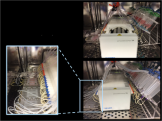
Tubule-on-a-chip comprised of tubes lined with either endothelial or epithelial cells grown together in a 3D channel
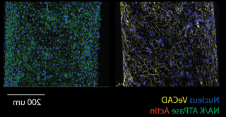
In situ vascular endothelial cells (left) and tubular epithelial cells (right), stained for: Nucleus (blue), VeCAD (yellow), NA/K ATPase (green), Actin (red)
One of the benefits of MPS models is that they can also incorporate biosensors that continuously measure genetic and proteomic signatures in response to specific stimuli. As MPS platforms permit many cell types and parameters to be analysed in ‘real time’ this allows increased throughput in terms of data generation, and provides results that are more translatable and relevant to data collected from patients than those obtained from previous 2D models.
So far there have been no reports of in vitro cellular models adequately mimicking kidney function. Patient-derived primary cells are desirable for modelling disease states in a laboratory setting and for testing potential drug candidates. However, these cells present challenges to scientists, including limited availability, loss of their physiological characteristics when in culture, and the fact that it is difficult to expand them to the scale needed for in vitro assays.
Miniorgans – another way to study kidney function
Organoids are a complementary approach for replicating organ function in the lab. Kidney organoids have been created that comprise at least 11 different cell types, which are needed for the full organ to function normally. Although the replicated structure is not identical to the actual organ, the mixture of cells creates interactions which emulate the situation seen in vivo.
Eventually, we hope that multiple kidney component-on-a-chip devices may be linked together to produce a whole organ-on-a-chip. This would create a uniquely translatable system with high fidelity to the whole human kidney.
Kidney-on-chip technology has the potential to enhance and accelerate our ability to translate science into innovative medicines for patients with CKD. By creating greater opportunities for sharing of expertise between different scientific and clinical disciplines – including engineering, biophysics, and biology – we will enhance our understanding of CKD and advance new approaches and technologies for treating kidney disease.